Saturday

Composite build up

CASE REPORT: A 41 year old male patient presented with the complaint of missing upper front teeth on examination upper left second incisor and canine were missing moreover upper left first incisor and first premolar had crown cutting done on it. Patient informed that he had bridge previously which was broken off  around 2 months back. Moreover patient upper left second premolar had class 2 extensive caries and not enough tooth structure was left. Patient had no interest in having any extra crown except upper front convetional bridge. Upper left second premolar had no history of pain or tender on percussion and it was vital. Because of these circumstances composite build up was decided to be done on upper left second premolar followed by which bridge work was completed with missing teeth.

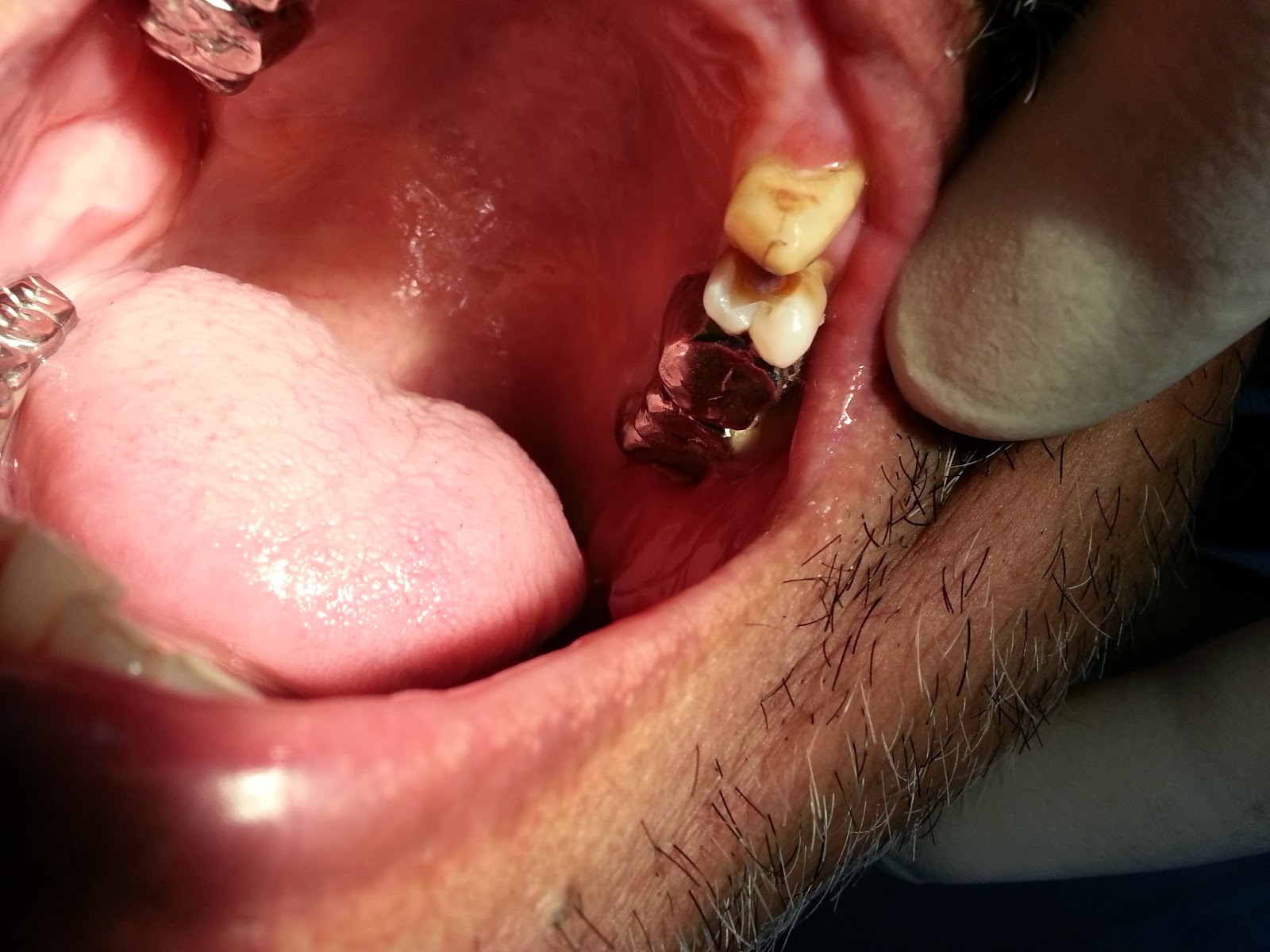
All the caries from upper left second premolar was removed and calcium hydroxide lining was placed around pulp. Followed by which composite build up was done with final polishing. A3 shade was choose on vita shade guide considering the bridge placement and opposite premolar on upper arch.

Image on the left shows upper left second premolar with completed treatment.

Vertical fracture

CASE REPORT: A 68 year old female patient presented with severe pain in her lower right back tooth since one day after biting on any hard object. On examination lower right first molar had vertical  fracture towards lingual side. the fracture line was extending below the gingiva and therefore prognosis of the lower right first molar was poor. Patient was wishing for tooth extraction.


The procedure was started after thorough patients history and radiograph. Area was anesthetized with inter dental nerve block along with buccal and lingual nerve block. small broken lingual fragment was removed followed by total tooth extraction. 


No fragments were left behind and there was no any soft tissue or alveolar bone damage.

Extraction of the patient on anticoagulant

CASE REPORT : A 57 year old male patient presented with grade 1 mobile right upper canine with severe continuous pain since last 4 days, on examination tooth was non vital and periapical radiolucency with respect to right upper canine was present. The tooth was tender on percussion.

On taking the medical history patient was on aspirin ( Disprin) 150 mg since last 4 years due to coronary heart disease. Patient was refereed to medical laboratory for bleeding time and clotting time which was prolonged. Patient's GP was consulted, emergency pulp extirpation was done on upper right canine and analgesics ( Paracetamol 500 mg 2 tab. every 6 hourly) given.

After 6 days BT and CT were measured again and they were normal therefore patient was called with full stomach and GP consultation.


Above images show complete extraction of upper right canine tooth with total control of post operative bleeding by adrenaline( Epinephrine 1:100000) containing 2 % Lidocaine HCL and pressure pack.

Chronic Generalised Periodontitis


CASE REPORT:
A 48 years old female patient presented in my clinic with severe pain in her gums since last 2 weeks on examination I had observed large quantity of calculus and poor oral hygiene. Upon taking the case history she revealed that she is diabetec and not taking care of herself for long. She had few grade 3 mobile teeth.


Patient was not regular attendee in dental clinic therefore DPT was taken to set up the base record and future treatment purposes which revealed marginal bone loss and calculus deposition there was no tenderness on percussion anywhere and no periapical pathologies were observed in DPT


First Visit : On first visit I removed all supragingival plaque and calculus under local anesthesia and motivate her to maintain proper oral hygiene, moreover I demonstrated right brushing technique and use of inter dental brushes in between the teeth. 0.2 % Chlorhexidine mouthwash was prescribed to use twice daily for 1 week


Second Visit(after 1 week): On second visit patient had shown sign of improvement, the swelling of marginal gingiva was subsided and I was able to remove subgingival calculus without any interuption by inflamed gums.


Third Visit(after 3 months): On third visit for regular check up patient was happy with appearance there was no pain or swelling on gingiva and she seemed motivated for regular dental check ups and maintaining good oral hygiene.


CLASS 4

CASE REPORT: A 47 year old male patient presented with broken upper left central incisor. patient had trauma around 6 years back since then incisor third of the left central incisor was missing.
On examination patient had no pain. tooth was vital and there was no any periapical pathology. fracture on left central incisor was involving enamel and dentin


Patient wished for more conservative and affordable treatment therefore composite build up was done with respect to upper left central incisor (CLASS 4)

Full ceramic crowns

CASE REPORT: A 47 year old male presented with the complain of pain and food lodgement between upper left second premolar (25) and upper left first molar (26). Patient had previous repeated filling done between the contact area and it had failed and patient had wished for crown placement with closed contacts between upper left second premolar and first molar.                                                                                                                                                               


 On examination there was a food lodgement broken composite filling on distal surface of the upper left second premolar and mesial surface of upper left first molar ( CLASS 2). Moreover there was a swelling around marginal gingiva with respect to both the teeth.
considering the poor oral hygiene and more consideration of self cleaning rather than aesthetic, supragingival margins of crown cutting were placed after patient's consent and full ceramic crowns were selected for placement.

proper depth and margins were  achieved with the help of putty index and occlusal clearance was maintained for full ceramic crowns following which putty and wash impression was taken with putty and light body rubber base impression material.

final crowns were placed with proper margins of the crowns and maintaining centric occlusion.


Friday

Treating one root canal

CASE REPORT: A 22 year old male patient presented with pain on his lower right back tooth since 3 weeks on examination lower right third molar tooth (46) was treated with root canal treatment 2 years back and no crown was provided.


Radiograph showed underfilled distal root canal with periapical radiolucency which was the reason for positive tenderness on percussion.


By keeping a young age of the patient in to the consideration total root canal treatment was avoided and hence only under filled root canal was exposed and treated
The estimated working length was determined of the distal root canal by apex locater but it lacked accuracy due to bio-mechanical preparation and apical flare of the root canal hence the proper working length of the distal root canal was determined with IOPA which is showed in the image below                                                                                                                                                                                             
The apical opening of the distal root canal was located at root resorption near periapical radiolucency


The distal root canal was obturated and later on tooth was crowned to provide additional strength. 

(apologies for the bad photograph as due to more transparency in radio-graphic film picture had to be taken in such a way.)


After completion of root canal treatment full ceramic crown was placed which is shown in the following pictures


Extra root canals

INTRODUCTION:

Knowledge of internal dental morphology is an extremely important in planning and administering endodontic therapy.The numerous anatomical variations existing in root canal system may contribute to the failure of root canal therapy. Failure to explore and instrument even one of the canals results in improper cleaning of root canal system and can lead to endodontic treatment failure. The mandibular first molar, the earliest permanent posterior tooth to be erupt, seems to be the tooth that most often requires root canal treatment. The usual canal distribution is two canals in the mesial root and one or two canals in the distal root.
CASE REPORT:
A 38 year-old female patient presented with a chief complaint of pain in the mandibular left posterior tooth for the past two weeks. Clinical examination revealed a carious left mandibular first molar (36). The clinical and radiographic findings led to a diagnosis of acute exaggeration of chronic periapical abscess.


Down below are the images and radiographs of given treatment.


Figure above shows Pre operative radiograph of the mandibular left first molar tooth showing the presence of more than one canal in distal root, in the patient in the case report

All canals were identified and working length was established by using apex locators and IOPAs


Below is the post operative x ray in which mesial orifices had an additional orifice between the two main buccal and lingual canals and distal orifices showing an additional orifice between two main buccal and lingual canals

Complete obturation was done with full ceramic crown and on recall after 2 months patient had no pain or any sign of infection.